Preclinical and clinical multimodality imaging, developed in the Labex IRON, integrates images acquired by different techniques: structural or anatomical images (CT, MRI) and functional images after injection of a radiopharmaceutical (PET, SPECT). Multimodality exploits this complementarity of images to enrich diagnostic information.
All the clinical imaging departments as well as most of the preclinical research centers of the Labex IRON (Angers, Caen, Nantes, Rennes, Tours, Toulouse) are equipped with multimodality imaging systems. In 2021, the Nantes University Hospital site acquired new equipment: a PET-MRI, the 5th French installation of this type. The Nantes region offers the only PET-MRI in the Grand-Ouest region used in clinical research.
β-imaging systems as well as optical imaging systems complete this network of imaging systems.
Since 2013, the IRON labex has had a platform (Imagys® workstation, Keosys) for collection, exchange between sites, and the archiving of qualified imaging data from preclinical research centers and clinical imaging services. This platform is shared between the sites of Nantes, Angers, Caen, Rennes, Toulouse and Tours.
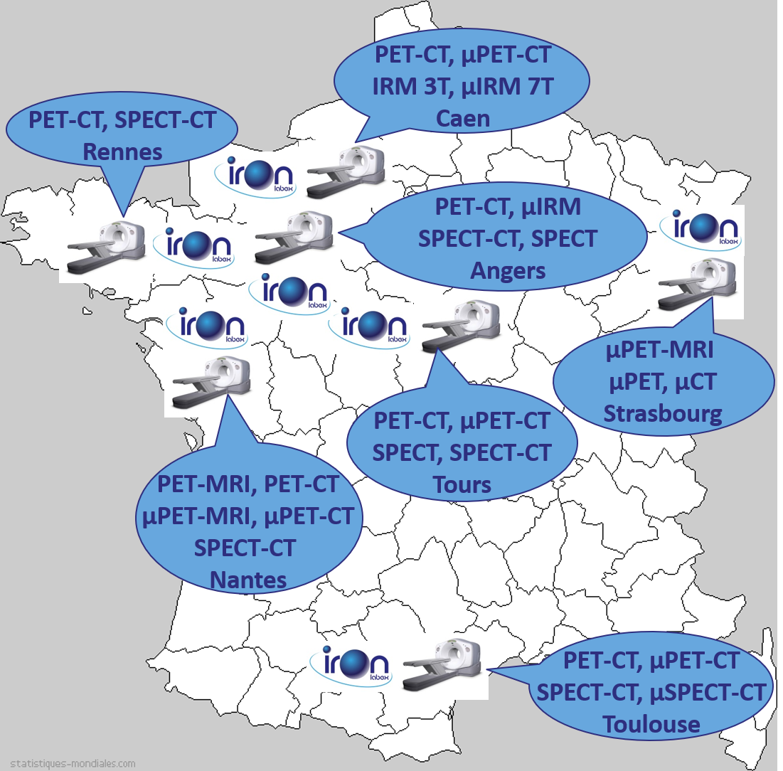
Imaging systems of the Labex IRON
Glossary:
CT (computarized tomography) = TDM (tomodensitométrie)
MRI (magnetic resonance imaging) = IRM (imagerie par résonance magnétique)
PET (positron emitting tomography) = TEP (tomographie par émission de positons)
SPECT (single photon emission computed tomography) = TEMP (tomographie d’émission monophotonique)